Anatomy and Function of the Meniscus

The meniscus is a C-shaped piece of cartilage that acts as a shock absorber and stabilizer within the knee joint. It is located between the femur (thigh bone) and the tibia (shin bone), and plays a crucial role in the proper functioning of the knee.
Location and Structure of the Meniscus
The meniscus is a crescent-shaped fibrocartilaginous structure that resides within the knee joint. There are two menisci in each knee: the medial meniscus, which is located on the inner side of the knee, and the lateral meniscus, which is located on the outer side of the knee. Both menisci are attached to the tibial plateau, the top surface of the tibia. The menisci are made up of tough, rubbery cartilage that helps to cushion the joint and distribute weight evenly across the knee.
Functions of the Meniscus
The meniscus plays several important roles in supporting the knee joint:
- Shock Absorption: The meniscus acts as a shock absorber, absorbing forces that are transmitted to the knee during activities like walking, running, and jumping. This helps to protect the articular cartilage, which is the smooth, slippery surface that covers the ends of the bones in the joint.
- Joint Stability: The meniscus helps to stabilize the knee joint by deepening the articular surface of the tibia, providing a better fit for the femur. This helps to prevent the femur from sliding off the tibia, especially during movements like twisting or pivoting.
- Load Distribution: The meniscus helps to distribute weight evenly across the knee joint, reducing the amount of stress on the articular cartilage. This is especially important during activities that put a lot of stress on the knee, such as running or jumping.
- Joint Lubrication: The meniscus helps to lubricate the knee joint by producing synovial fluid, which helps to reduce friction between the joint surfaces.
Types of Menisci
The two menisci in the knee have slightly different shapes and functions:
- Medial Meniscus: The medial meniscus is C-shaped and is more firmly attached to the tibial plateau than the lateral meniscus. This makes it more susceptible to tears, particularly during activities that involve twisting or pivoting movements.
- Lateral Meniscus: The lateral meniscus is more O-shaped and is less firmly attached to the tibial plateau than the medial meniscus. This makes it less susceptible to tears, but it can still be injured during high-impact activities.
Causes and Types of Meniscus Tears

A meniscus tear is a common injury that occurs when the cartilage in the knee is torn. This cartilage acts as a shock absorber and helps to distribute weight evenly across the knee joint. Tears can range from small, insignificant ones to large, complex ones that can significantly impact knee function.
Causes of Meniscus Tears
Meniscus tears can occur due to various factors, including sports injuries, degenerative changes, and trauma.
- Sports Injuries: Meniscus tears are frequently seen in athletes who participate in high-impact sports like football, basketball, and skiing. These activities often involve twisting or pivoting movements that can stress the knee joint, leading to a tear.
- Degenerative Changes: As we age, the meniscus naturally deteriorates, becoming thinner and weaker. This process, known as degeneration, can make the meniscus more susceptible to tears, even with minimal trauma.
- Trauma: A direct blow to the knee, such as a car accident or a fall, can also cause a meniscus tear.
Types of Meniscus Tears
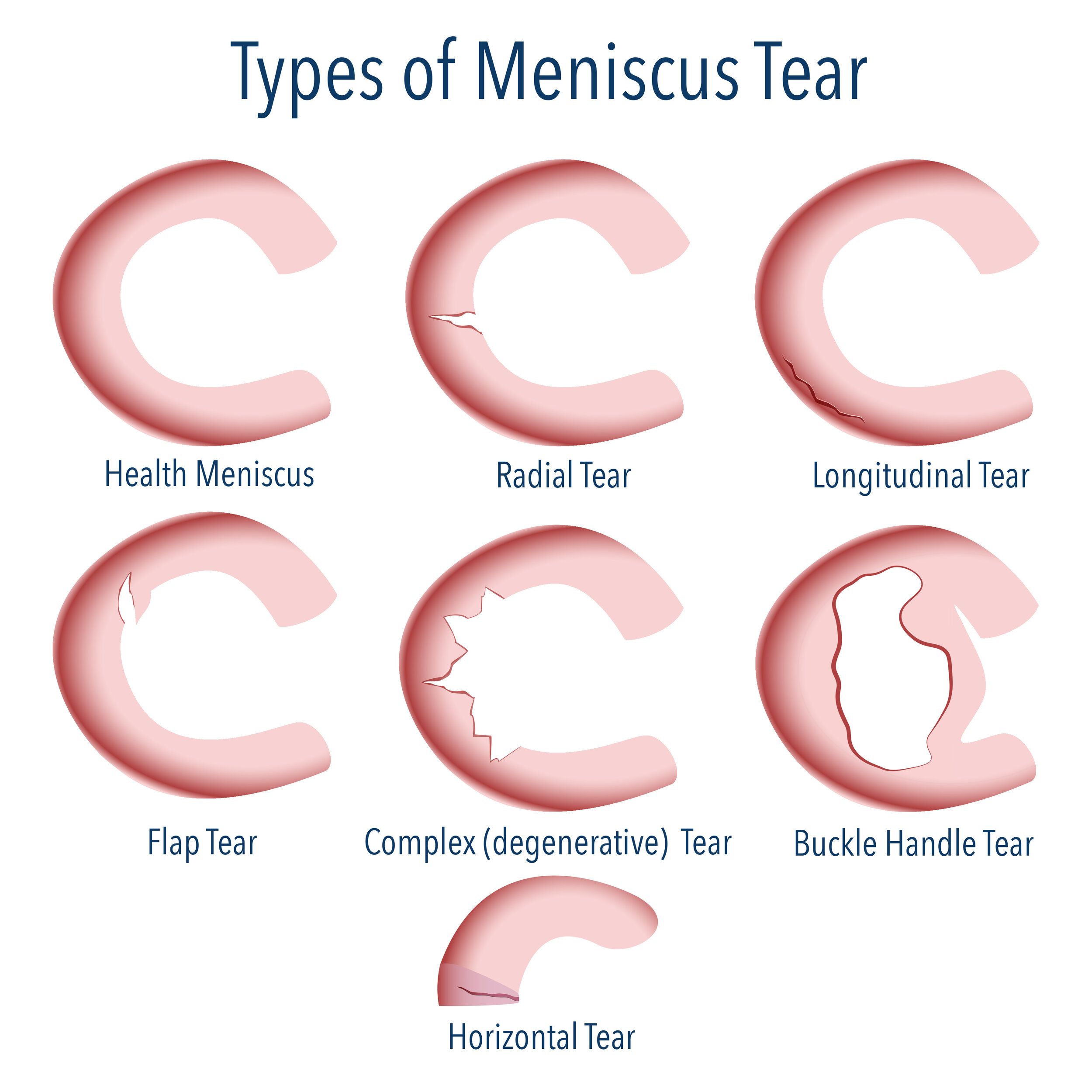
Meniscus tears are classified based on their location, shape, and severity.
- Longitudinal Tears: These tears run lengthwise along the meniscus. They are often caused by a twisting or pivoting motion and can vary in size and severity.
- Radial Tears: These tears run from the outer edge of the meniscus toward the center. They are often caused by a direct blow to the knee or a sudden compression of the joint.
- Flap Tears: These tears involve a piece of the meniscus being torn off and left as a loose flap. They can cause significant pain and instability in the knee.
Factors Influencing Severity and Symptoms
The severity and symptoms of a meniscus tear depend on various factors:
- Location of the Tear: Tears in the outer portion of the meniscus, which has a better blood supply, tend to heal better than tears in the inner portion, which has a limited blood supply.
- Size and Shape of the Tear: Larger tears and complex tears are more likely to cause significant pain and instability.
- Age and Activity Level: Younger, more active individuals may experience more severe symptoms and require more aggressive treatment.
Symptoms and Diagnosis of Meniscus Tears

A meniscus tear can cause a variety of symptoms, ranging from mild discomfort to severe pain and disability. Understanding these symptoms is crucial for early diagnosis and appropriate treatment. This section will discuss the common symptoms associated with meniscus tears and the diagnostic methods used to confirm the injury.
Symptoms of Meniscus Tears
Meniscus tears can manifest in various ways, depending on the severity and location of the tear. The most common symptoms include:
- Pain: This is often the first and most prominent symptom. Pain may be localized to the knee joint, particularly around the affected meniscus, and may worsen with activities such as squatting, twisting, or climbing stairs.
- Swelling: Swelling in the knee joint is another common symptom, often developing shortly after the injury or within a few hours.
- Locking: A feeling of the knee locking or catching may occur if a piece of the torn meniscus becomes trapped in the joint space, preventing the knee from extending fully.
- Instability: The knee may feel unstable or give way, particularly during activities that require twisting or pivoting movements.
- Stiffness: The knee may feel stiff and limited in range of motion, especially after periods of rest or inactivity.
- Clicking or Popping: Some individuals may experience a clicking or popping sensation in the knee joint, especially during movement.
Diagnosis of Meniscus Tears
Diagnosing a meniscus tear typically involves a combination of physical examination, imaging studies, and, in some cases, arthroscopy.
Physical Examination
A thorough physical examination is the first step in diagnosing a meniscus tear. The doctor will assess the knee joint for pain, swelling, tenderness, and range of motion. They may also perform specific tests to evaluate the stability of the knee and check for signs of a meniscus tear, such as:
- McMurray’s Test: This test involves flexing and extending the knee while rotating the leg. A clicking or popping sensation during this maneuver can suggest a meniscus tear.
- Apley’s Compression Test: This test involves applying pressure to the knee while rotating the leg. Pain during this test can indicate a meniscus tear.
- Lachman’s Test: This test assesses the stability of the anterior cruciate ligament (ACL), which is often injured in conjunction with a meniscus tear.
Imaging Studies
Imaging studies, such as magnetic resonance imaging (MRI), are often used to confirm the diagnosis and determine the extent of the tear.
- Magnetic Resonance Imaging (MRI): MRI is a highly sensitive imaging technique that provides detailed images of the knee joint, including the meniscus. It can clearly visualize tears in the meniscus and help determine the location, size, and severity of the tear.
Arthroscopy
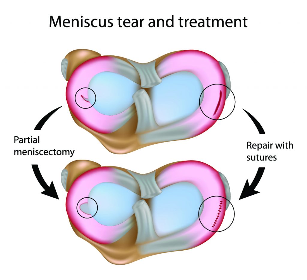
Arthroscopy is a minimally invasive surgical procedure that allows the doctor to visualize the inside of the knee joint. It is often performed to confirm the diagnosis, assess the extent of the tear, and repair or remove the damaged meniscus.
Comparison of Diagnostic Techniques
Each diagnostic technique has its own strengths and limitations.
| Technique | Strengths | Limitations |
|---|---|---|
| Physical Examination | Non-invasive, relatively inexpensive, can provide valuable information about the knee joint | May not be able to definitively diagnose a meniscus tear, particularly in cases with subtle symptoms |
| MRI | Highly sensitive and specific for diagnosing meniscus tears, can provide detailed information about the location, size, and severity of the tear | Expensive, not always readily available, may not be necessary in all cases |
| Arthroscopy | Allows direct visualization of the knee joint, can be used to both diagnose and treat meniscus tears | Invasive procedure, requires anesthesia, carries risks associated with any surgery |
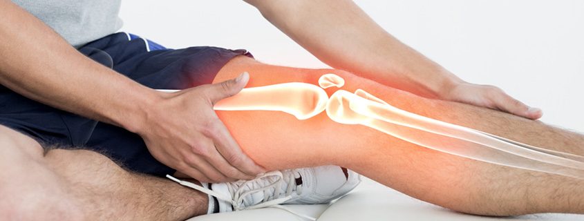
Meniscus tear – The meniscus, a C-shaped cartilage in the knee, acts as a shock absorber and helps stabilize the joint. Tears in the meniscus, often caused by sudden twisting or impact, can lead to pain, swelling, and limited mobility. A recent example of such an injury is the jj mccarthy knee injury , highlighting the impact such injuries can have on athletes.
Understanding the structure and function of the meniscus is crucial for preventing and managing these common knee injuries.
A meniscus tear, a cruel twist of fate for any athlete, can be a devastating setback. The sharp pain, the sudden loss of mobility – it’s a nightmare. Imagine the agony, the frustration, the fear of the unknown. For Jahmyr Gibbs, a rising star in the NFL, a meniscus tear could be a career-altering event.
Jahmyr Gibbs , a beacon of hope for his team, now faces a long road to recovery. A meniscus tear is a harsh reminder of the fragility of even the most powerful athletic bodies.
